
General Oral & Maxillofacial Surgery
Cysts & Tumors
Surgical treatment of cysts and benign or malignant tumors of the jaws and oral cavity.
Overview
Surgical treatment of cysts and benign or malignant tumors of the jaws and oral cavity.
Every recommendation follows an individual assessment of your concerns, anatomy and goals. The sections below explain how this treatment is typically diagnosed, planned and carried out.
Who May Benefit
- Patients referred by a dentist, orthodontist or physician for specialist surgical care
- Patients with injuries, cysts, tumors or other conditions of the jaws and face
- Patients requiring reconstruction following trauma or disease
Conditions Treated
- Jaw cysts
- Benign odontogenic tumors
- Salivary gland lesions
Diagnosis
Diagnosis is based on clinical examination and appropriate imaging, often CBCT or CT, and where needed a biopsy to confirm the nature of a lesion.
Treatment Planning
Plans are tailored to the diagnosis and may involve collaboration with other specialists. The balance between treating the condition and preserving function and appearance is central to planning.
Virtual Planning
For cysts and tumors of the jaws, 3D imaging maps the lesion and its relationship to nerves, teeth and the sinus. Planning sets safe removal margins and, when part of the jaw must be removed, designs the reconstruction in advance with patient-specific guides and plates.
Surgical Overview
Surgical approach depends on the diagnosis and may involve enucleation, resection and, where needed, reconstruction.
Recovery
Recovery ranges from a few days after routine minor oral surgery to several weeks after major reconstructive surgery. Follow-up may include monitoring imaging.
Risks & Considerations
All surgery carries risk. Potential risks are discussed in detail during consultation, and may include:
- Swelling and discomfort
- Bleeding or infection
- Altered sensation
- Recurrence of some lesions
- Risks associated with anaesthesia
Frequently Asked Questions
Do I need a referral?
Referral requirements depend on your location and healthcare system. Patients and referring clinicians are welcome to use the consultation form to discuss next steps.
Cases from the clinic
Real cases treated by Dr. Bakri, published with patient consent.
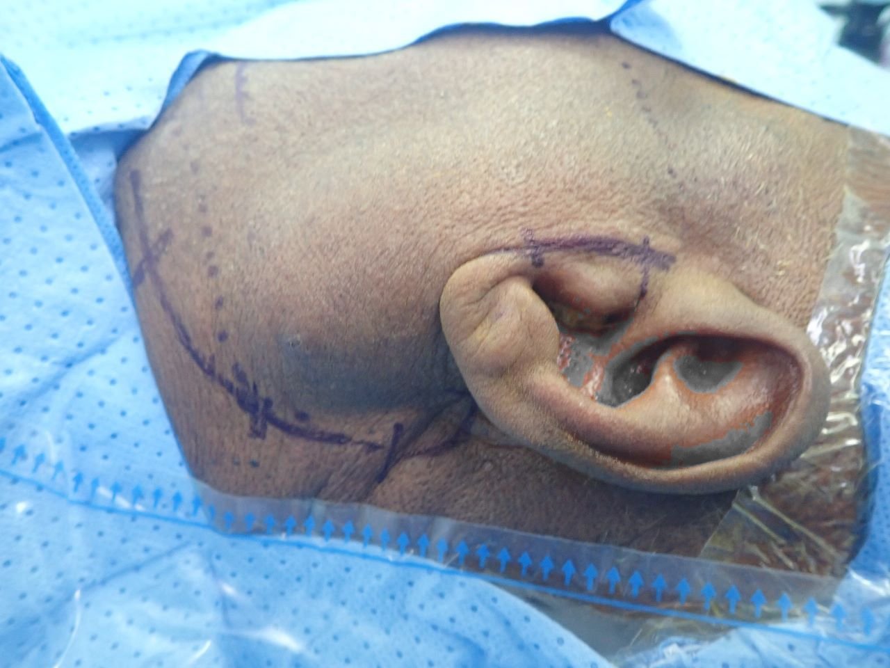
Before Treatment 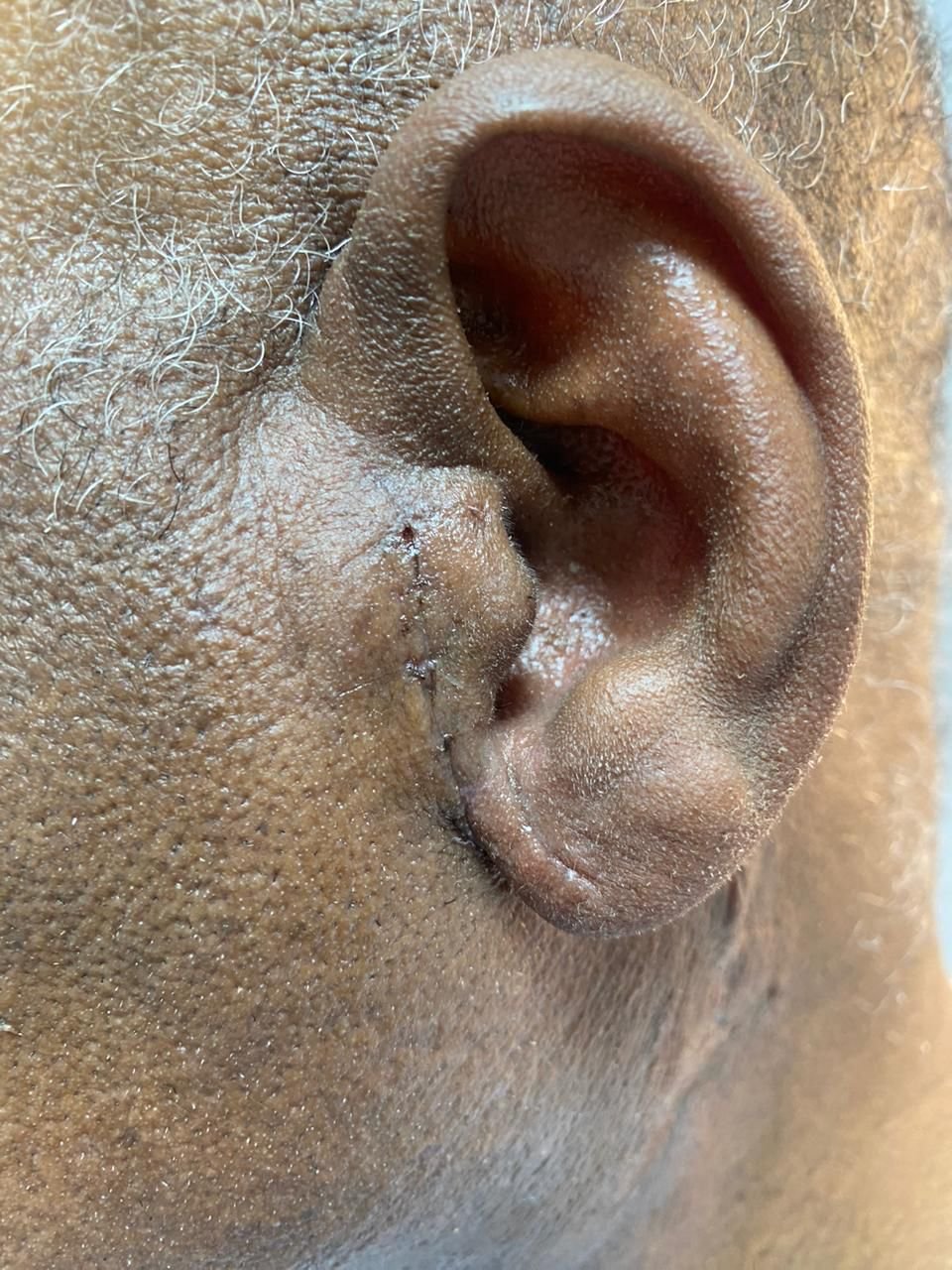
After Treatment Cysts, tumors & salivary glandsParotid tumor removed through an incision hidden in the ear crease
A swelling in front of and below the ear. The incision followed the natural crease in front of the ear, so after healing it blends into the ear's contours.
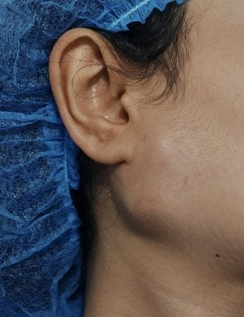
Before Treatment 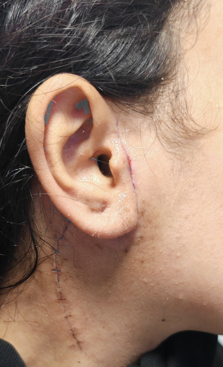
After Treatment Cysts, tumors & salivary glandsParotid tumor: an incision that follows the ear and neck creases
The tumor was mapped precisely with MRI, then the incision was placed in front of the ear and along natural neck creases for safe access and a discreet scar.
Before Treatment 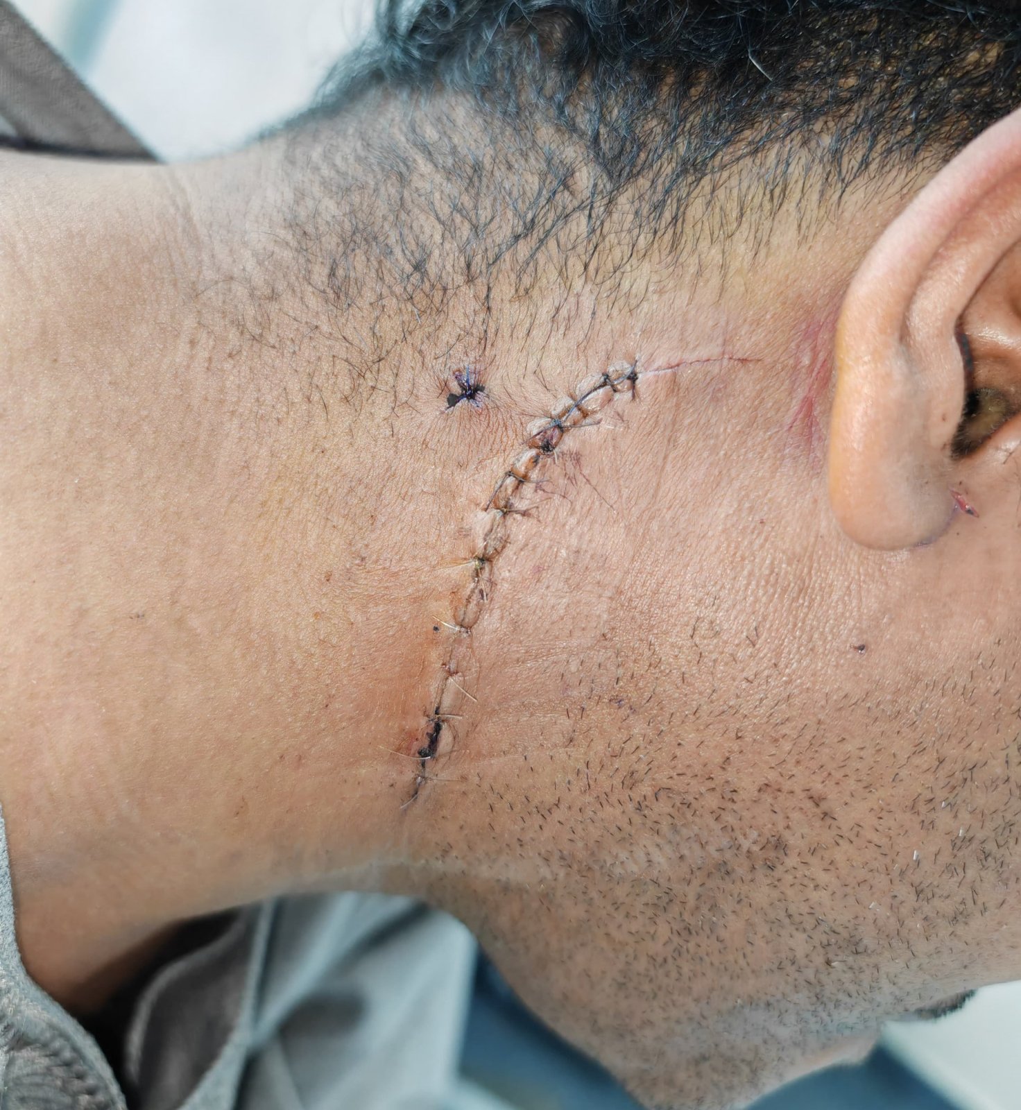
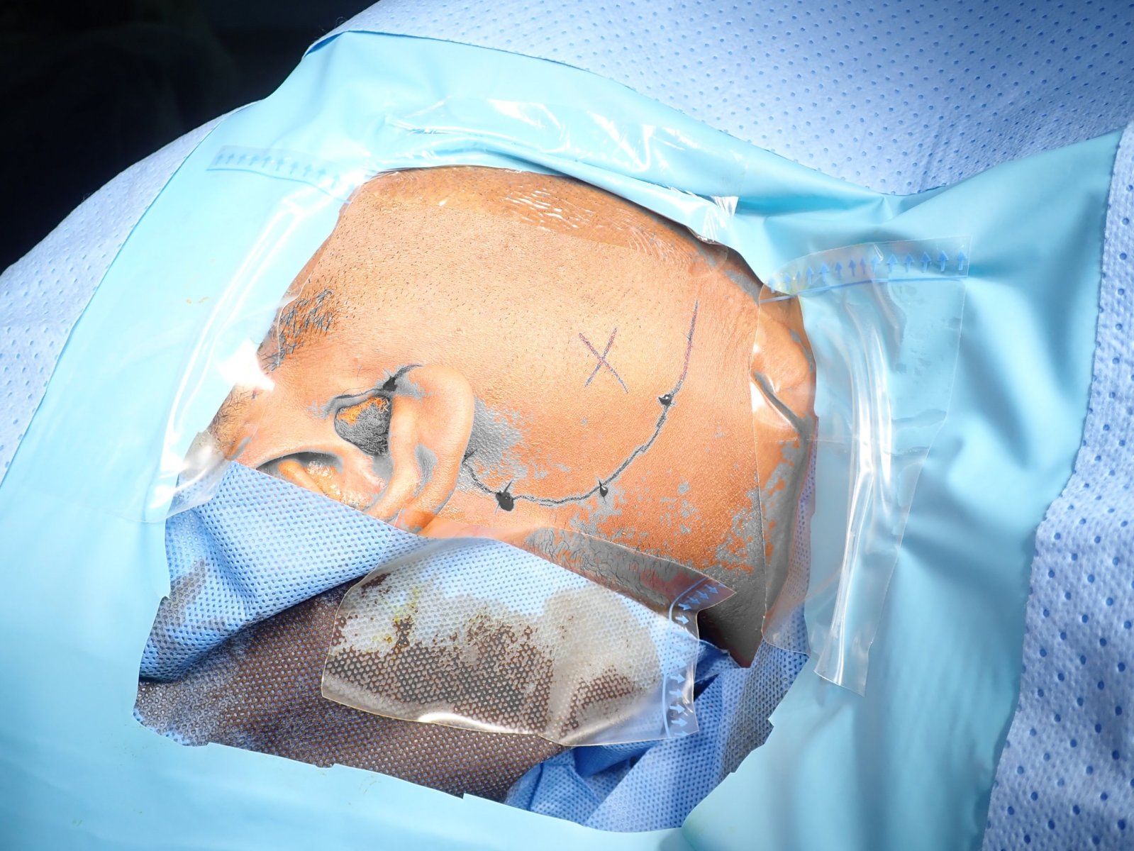
After Treatment Cysts, tumors & salivary glandsParotid tumor: an incision behind the ear, covered by the hair
The incision was planned behind the ear and within the hairline, with no visible cut on the cheek, so the hair covers the scar once healed.
Discuss your options with Dr. Bakri
A consultation is the first step toward an accurate diagnosis and an individual plan.